| |||||||||||||||||||
 |
||
|
||
 Users Online
Forum Threads
Newest Threads
· Tachinidae ID· Tephritidae ID · Family ID => Simulium · Rhamphomyia? · Tachinidae -> Leucos... Hottest Threads
Theme Switcher
Last Seen Users
Latest Photo Additions
|
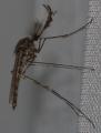
More Information about: ventral
Comments
Post Comment
Please Login to Post a Comment.
| Date and time
Login
Temporary email?
Due to fact this site has functionality making use of your email address, any registration using a temporary email address will be rejected.
Paul Donate
Latest Articles
· Voltinism in Chaoborus
· Vitaliy Nikolaevich ... · Mating behaviour and... · Oviposition into the... · African Invertebrates Syrph the Net
License Agreement - Click to Download Public files of Syrph the Net can be downloaded HERE Last updated: 25.08.2011 Shoutbox
You must login to post a message. 23.06.25 18:10 If you have some spare money, there is a copy (together with keys to pupae and larvae) for sale by Hermann L. Strack, Loguivy Plougras, France
23.06.25 11:18 Appreciate it, Tony Irwin! I got the hint to use the key next to Langton and Pinder key for females of Chironomidae. So no specific queries, except the keys...
I will keep this on my list and hope th
19.06.25 15:33 I have the hard copy book, if you have any specific queries, but I'm not scanning the 500+ pages!
02.06.25 18:26 Anyone has "Chironomidae of the Holarctic region. Keys and diagnoses. Part 3. Adult Males Entomologica Scandinavica Supplement 34"?
smolwaarneming@gma
il.com 28.05.25 20:57 I have Russian Coenosia.
nikita6510@ya.ru
28.05.25 12:25 Is someone able to share with me "A key to the Russian species of the genus Coenosia"?
08.05.25 18:22 I have
03.05.25 08:35 Does someone has a scan of Nartshuk E.P. 2003. Key to families of Diptera (Insecta) of the fauna of Russian and adjacent countries. Proceedings of the Zoological Institute Vol. 294: 1-252 for me?
10.03.25 18:02 We are looking for a new webmaster
https://diptera.in
fo/forum/viewthrea d.php?thread_id=11 5023&rowstart=20 04.03.25 17:10 Please use the link posted below to remember and honour Paul, if you wish
|
 which is raised on one side if the seta is slanted. Setae are usually connected to nerves and have a sensory function.<br /><img src='../../infusions//terms/images/no_image.gif' style='vertical-align:middle;' />] delay=[0] fade=[on]")
|
images in Diptera Gallery and Forum of their respective owners Powered by PHP-Fusion copyright © 2002 - 2025 by Nick Jones. Released as free software without warranties under GNU Affero GPL v3. SimpleAsThat |
||
| Render time: 0.21 seconds | 229,979,740 unique visits | ||
(outside) of a transverse section is then divided into eight imaginary parts, which (looking down the leg towards the body read in the direction of a clock)
dorsal
postero-dorsal
posterior
postero-ventral
ventral
antero-ventral
anterior
antero-dorsal